X-rays, including Digital X-rays and 3D CBCT Imaging, are indispensable tools in modern diagnostic practices, playing a crucial role in the accurate detection of various dental conditions. These imaging techniques enable dental professionals to examine the internal structures of the teeth, bones, and surrounding tissues, facilitating early detection of potential issues and the development of effective treatment plans. Digital X-rays have revolutionized diagnostic imaging by reducing radiation exposure and enhancing image clarity, while 3D CBCT (Cone Beam Computed Tomography) Imaging offers a three-dimensional view that significantly improves the precision of diagnoses, especially for complex dental conditions. As technology advances, these imaging systems continue to enhance the capability of clinicians to provide more accurate and efficient care.
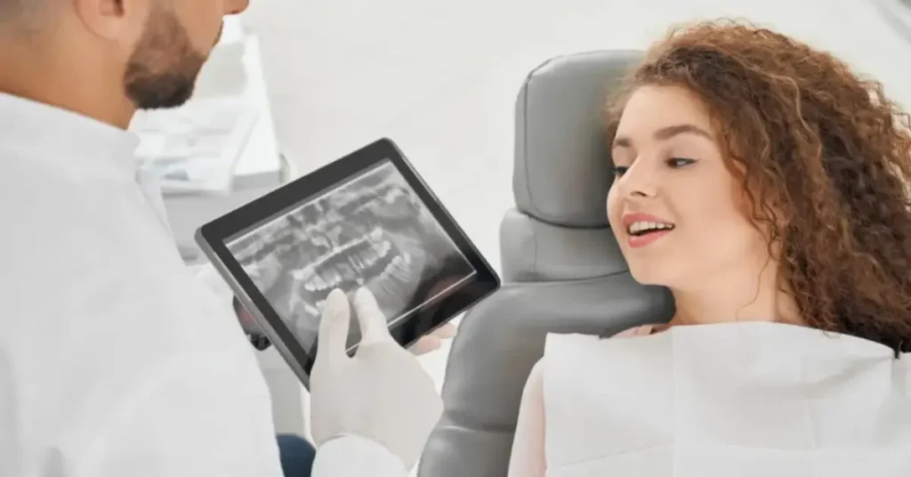
Digital X-rays
Digital X-rays are a significant advancement in diagnostic imaging, replacing traditional film-based X-rays. By using digital sensors, they provide clearer, more detailed images that can be immediately analyzed on a computer. One of the main benefits is the reduction in radiation exposure compared to conventional X-ray systems. Digital X-rays offer a quicker turnaround time, allowing dental professionals to immediately assess images for conditions such as cavities, infections, and bone loss.
- Reduced Radiation Exposure: Digital X-rays utilize a significantly lower level of radiation compared to traditional film-based X-rays. This reduction in radiation ensures better safety for patients, especially for those who require frequent imaging, without compromising on the clarity or quality of the diagnostic image.
- Instant Image Availability: With digital sensors, the images are available almost immediately on a computer screen. This reduces waiting time, allowing dental professionals to quickly review and diagnose the images, which in turn speeds up the entire process, from diagnosis to treatment planning, making the experience more efficient for the patient.
- Enhanced Image Clarity: Digital X-rays capture higher resolution images, allowing for greater detail and precision when examining dental structures. This enhanced clarity helps to detect even the smallest dental issues, such as early-stage cavities or fractures, that might otherwise go unnoticed with traditional X-ray methods.
3D CBCT Imaging
3D CBCT Imaging is an advanced imaging technique that captures detailed, three-dimensional images of the dental structures, including the teeth, jaws, and surrounding tissues. Unlike traditional X-rays, which only provide two-dimensional images, CBCT offers a comprehensive view, allowing dental professionals to assess the volume and density of bone, detect hidden infections, and evaluate the alignment of the teeth and jaw. This imaging method is particularly beneficial for planning procedures such as dental implants, orthodontics, and complex surgeries.
- Comprehensive View: The three-dimensional perspective provided by CBCT imaging allows dental professionals to examine a patient’s entire oral structure, including soft and hard tissues. This comprehensive view is invaluable for planning complex procedures, such as dental implant placement or corrective jaw surgery, where detailed spatial relationships between teeth, bone, and surrounding tissues must be considered.
- Precision in Diagnosis: 3D CBCT imaging offers unparalleled diagnostic accuracy by capturing detailed images of the teeth, jawbones, and surrounding structures. This level of precision helps identify conditions that would otherwise be missed with traditional X-rays, such as hidden infections or structural abnormalities that could affect treatment planning or outcomes.
- Detailed Treatment Planning: With the ability to visualize dental structures in three dimensions, dental professionals can plan treatments, such as implants or orthodontic procedures, with a higher degree of accuracy. This allows for better prediction of treatment success, reduces the likelihood of complications, and improves patient satisfaction by ensuring that treatments are tailored to their specific needs.
Advanced Diagnostics
Advanced diagnostic tools, such as Digital X-rays and 3D CBCT Imaging, have revolutionized the field of dentistry. These technologies allow for early detection of various dental issues, from cavities to tumors, by providing a clear and accurate picture of the oral structures. The increased precision of these diagnostic methods has led to better treatment outcomes, as it allows dental professionals to identify problems before they become severe, reducing the need for invasive procedures.
- Early Detection of Dental Issues: Advanced imaging techniques allow for the detection of dental problems at the earliest possible stage, which is crucial in preventing the progression of conditions like tooth decay, gum disease, or even oral cancers. Early detection not only saves teeth but also reduces the need for more invasive and costly treatments down the line.
- Comprehensive Analysis: The ability to examine dental structures from multiple angles—whether through 2D digital images or 3D CBCT scans—provides dental professionals with a thorough understanding of the patient’s condition. This comprehensive analysis ensures that all aspects of a dental issue are identified, leading to more accurate diagnoses and more effective treatment plans.
- Minimizing Invasive Procedures: By identifying issues early and accurately, advanced diagnostics help minimize the need for invasive treatments. Conditions that could have required surgery or complex interventions can often be treated with more conservative methods, leading to faster recovery times and less discomfort for patients.
Precision Treatment
Precision treatment is one of the major benefits of advanced imaging technologies such as Digital X-rays and 3D CBCT Imaging. With a clear understanding of the patient’s anatomy, dental professionals can tailor treatments to meet the individual needs of each patient. This level of precision ensures that procedures like dental implants, orthodontic work, and restorative treatments are carried out with the utmost accuracy, leading to more successful and lasting results.
- Tailored Treatment Plans: By providing clear, detailed images of the patient’s teeth, bones, and surrounding tissues, advanced imaging allows dental professionals to design customized treatment plans. These personalized plans address the specific needs of the patient, ensuring the most effective and appropriate treatments are chosen for optimal outcomes.
- Reduced Treatment Times: When dental professionals have access to highly accurate, detailed images, they can plan and execute procedures with greater precision. This leads to more efficient treatments, as there is less need for guesswork or adjustments during the procedure. In many cases, patients may experience shorter treatment times and fewer visits.
- Improved Long-Term Outcomes: Precision in treatment planning and execution often translates into improved long-term results for patients. By addressing the issue accurately from the start, dental professionals can ensure that the treatment is effective, durable, and minimizes the risk of complications in the future, leading to higher patient satisfaction and better overall health outcomes.
X-ray Technology
X-ray technology is the backbone of modern dental diagnostics, providing an essential tool for visualizing internal structures that are otherwise hidden from view. Digital X-rays and 3D CBCT Imaging have greatly advanced traditional X-ray methods, making them more efficient and effective in diagnosing various dental and oral health issues. Digital X-rays, in particular, allow for quick image capture and instant access, while CBCT takes X-ray technology to the next level by offering detailed 3D images of the teeth, jawbones, and surrounding tissues. These advancements in X-ray technology have led to significant improvements in treatment planning and patient care.
- Digital Imaging Efficiency: Digital X-rays replace traditional film, eliminating the need for time-consuming film development. This not only saves time but also reduces costs, while providing higher-quality images that are easier to store and retrieve, enhancing the workflow and efficiency of dental practices.
- Advanced 3D Imaging: CBCT imaging is a breakthrough in X-ray technology, offering a detailed, three-dimensional perspective of the oral cavity. This advanced imaging method allows for more precise planning and evaluation of dental issues, especially in complex cases such as implants, root canals, and jaw surgeries.
- Enhanced Diagnostic Capabilities: X-ray technologies like digital and CBCT imaging provide a much clearer, more comprehensive view of a patient’s dental health compared to older methods. This results in more accurate diagnoses, fewer missed conditions, and better treatment outcomes, helping to ensure that patients receive the best possible care.
Conclusion
Advancements in diagnostic technologies like Digital X-rays and 3D CBCT Imaging have significantly transformed how dental professionals approach diagnosis and treatment planning. These cutting-edge tools offer a clearer, more detailed view of a patient’s oral health, enabling the early detection of dental issues, precise treatment planning, and improved long-term outcomes. At Rayen’s Dental Clinic, we are dedicated to utilizing these state-of-the-art technologies to provide our patients with the most effective, personalized care, ensuring each individual receives the highest standard of dental treatment.
Read also: Ultrasonic Cleaning


